Tendon Diagram Labeled - Knee Wikipedia : Read over the labeled diagram carefully, then switch to a blank copy of the same diagram and try to fill in the names of as many muscles as you can remember.
Tendon Diagram Labeled - Knee Wikipedia : Read over the labeled diagram carefully, then switch to a blank copy of the same diagram and try to fill in the names of as many muscles as you can remember.. Huesos del miembro superior arm anatomy arm bones human anatomy Posted in diagrams , muscles | tagged human muscles , human muscles anatomy , muscle , muscle chart , muscle diagram , muscles , muscles anatomy. The knee joint, you need a perfectly labeled diagram of the knee. Visceral muscle is found inside of organs like the stomach, intestines, and blood vessels. On the other hand, the insertion is where a tendon attaches that muscle to the *more* movable bone.
Tendon diagram simple / 8.4c: However, the long head of the biceps brachii is one of the more common tendons to rupture. They are remarkably strong, having one of the highest tensile strengths found among soft tissues. A complete rupture of any tendon in the body is rare. Riesige auswahl an cds, vinyl und mp3s.
The Knee Anatomy Injuries Treatment And Rehabilitation from cdn-prod.medicalnewstoday.com Shoulder tendons chart ~ labeled anatomy chart of shoulder ligaments on white background stocktrek images. This diagram depicts human anatomy tendons and ligaments.human anatomy diagrams show internal organs, cells, systems, conditions, symptoms and sickness information and/or tips for healthy living. Tendon diagram simple / 8.4c: Skeletal muscle diagram muscle fascia heart development types muscles fascia human body muscle and fascia heart cell fascia skeletal muscle cell anatomy muscular contraction. Label a diagram if you're a visual learner. A muscle's origin is where a tendon attaches it to the *less* movable bone. A tendon is a structure that connects muscle to bone, and the biceps are connected by tendons at both the elbow and shoulder joints. Spend some time revising this diagram by connecting the name and location of each structure with what you've just learned in the video.
They cause motion and produce a force that the body uses to move and manipulate the body.
See muscle contraction diagram stock video clips. Read over the labeled diagram carefully, then switch to a blank copy of the same diagram and try to fill in the names of as many muscles as you can remember. As these muscles contract and relax, they move skeletal bones to create movement of the body. Skeletal muscle diagram muscle fascia heart development types muscles fascia human body muscle and fascia heart cell fascia skeletal muscle cell anatomy muscular contraction. This will help you to understand the mechanism as well as the working. Branches of the femoral artery supply. Superficial and deep anterior muscles of upper body superficial and deep posterior muscles of upper body. Link to pt program exercise templates. In human anatomy, the peroneus longus (also known as fibularis longus) is a superficial muscle in the lateral compartment of the leg, and acts to evert and plantarflex the ankle. Arm muscle diagram labeled simple : Make writing personal training programs easy with these custom designed exercise templates, and keep your clients focused and progressing. The knee joint, you need a perfectly labeled diagram of the knee. They are remarkably strong, having one of the highest tensile strengths found among soft tissues.
Labeled pectoralis transversus, and the pectoantebrachialis is labeled pectoralis descendens. Tears of the achilles tendon can be tiny (microtears), or large, causing pain, swelling, and impaired movement. The subacromial bursa lies between the rotator cuff and shoulder blade and protects the tendons in this area. The knee is one of the largest and most complex joints in the body. Muscle tone is a natural condition in which a skeletal muscle stays partially contracted at all times.
Amazon Com Labeled Anatomy Chart Of Full Body Male Black Float Frame Canvas Art Artwork Posters Prints from m.media-amazon.com It runs down the back of the lower leg and connects the calf muscle to the. If you would like to learn all the parts of the foot structure, you have come to the right place. Tears of the achilles tendon can be tiny (microtears), or large, causing pain, swelling, and impaired movement. Visceral muscle is found inside of organs like the stomach, intestines, and blood vessels. Tendon diagram simple / 8.4c: The patellar tendon attaches the patella to the top of the tibia. Rupture of the biceps tendon. Diagram of tendons in forearm 👉 we are pleased to provide you with the picture named right arm muscle and tendon anatomywe hope this picture right arm muscle and tendon anatomy can help you study and research.
The weakest of all muscle tissues, visceral muscle makes organs contract to move substances through the organ.
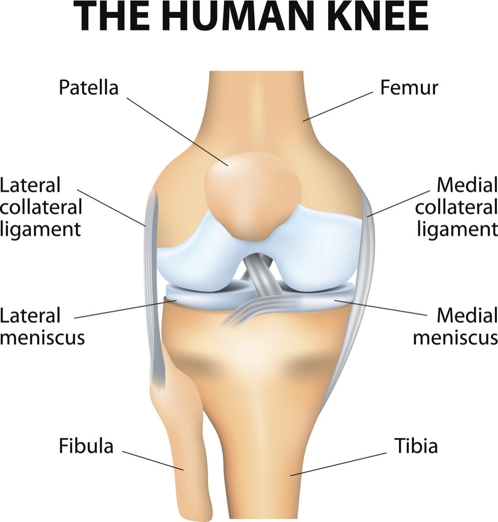
To understand one of the most complex joints of our body i.e. The only bone in the thigh is the femur, which extends from the hip to the knee.it can resist forces of 1,800 to 2,500 pounds, so it is not easily fractured. Learn about the anatomy and physiology of tendons. The subacromial bursa lies between the rotator cuff and shoulder blade and protects the tendons in this area. The knee joint, you need a perfectly labeled diagram of the knee. Link to pt program exercise templates. A labeled diagram of the knee with an insight into its working. In the diagram of the humerus this structure receives the head of the radius when the forearm is flexed. It is controlled by the obturator nerve. Muscle tone is a natural condition in which a skeletal muscle stays partially contracted at all times. System diagram labeled 209 human muscular system diagram labeled : Related posts of muscles and tendons of the leg muscle anatomy head. Shoulder tendons chart ~ labeled anatomy chart of shoulder ligaments on white background stocktrek images.
Related posts of muscles and tendons of the leg muscle anatomy head. Superficial and deep anterior muscles of upper body superficial and deep posterior muscles of upper body. Tendon diagram simple / 8.4c: In human anatomy, the peroneus longus (also known as fibularis longus) is a superficial muscle in the lateral compartment of the leg, and acts to evert and plantarflex the ankle. Muscle charts of the human body for.
Tendon Diagram Labeled Synovial Joint Diagram Labeled Anatomy Chart With Two Related Online Courses On Physioplus from i.pinimg.com Visceral muscle is found inside of organs like the stomach, intestines, and blood vessels. Labeled pectoralis transversus, and the pectoantebrachialis is labeled pectoralis descendens. The only bone in the thigh is the femur, which extends from the hip to the knee.it can resist forces of 1,800 to 2,500 pounds, so it is not easily fractured. Your biceps tendons attach the biceps muscle to bones in the shoulder and in the elbow. The patellar tendon attaches the patella to the top of the tibia. The fleshy, thick part of the muscle is called its belly. 17 photos of the diagram of shoulder muscles and tendons. Read over the labeled diagram carefully, then switch to a blank copy of the same diagram and try to fill in the names of as many muscles as you can remember.
Read over the labeled diagram carefully, then switch to a blank copy of the same diagram and try to fill in the names of as many muscles as you can remember.
They cause motion and produce a force that the body uses to move and manipulate the body. Muscle anatomy head 12 photos of the muscle anatomy head dog head muscle anatomy, human. In human anatomy, the peroneus longus (also known as fibularis longus) is a superficial muscle in the lateral compartment of the leg, and acts to evert and plantarflex the ankle. Achilles tendon the achilles tendon is a band of tissue that connects a muscle to a bone. This diagram depicts muscle in the body 744×1054 with parts and labels. Muscle charts of the human body for. If you tear the biceps tendon at the shoulder, you may lose some strength in your arm and have pain when you forcefully turn your arm from palm down to palm up. Rupture of the biceps tendon. This will help you to understand the mechanism as well as the working. Posted in diagrams , muscles | tagged human muscles , human muscles anatomy , muscle , muscle chart , muscle diagram , muscles , muscles anatomy. A labeled diagram of the knee with an insight into its working. Tendons attach muscles to bones. Arm muscle diagram labeled simple :


0 Komentar